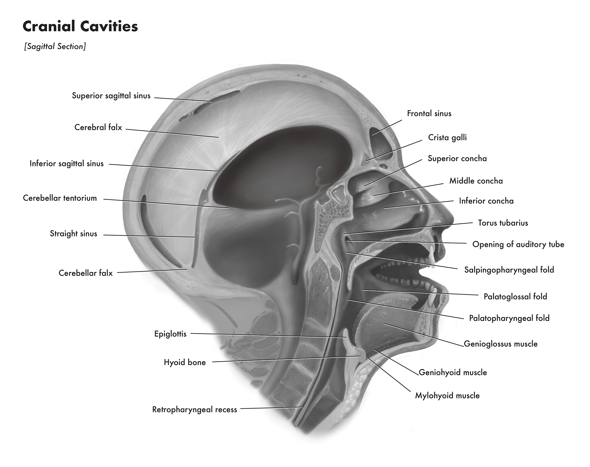
Cranial Cavities
The purpose of this illustration was to create an educational piece that shows
the internal anatomy of the human head. Specifically focusing on the cavities. A real perserved human
specimen was used as reference.
Continue reading to learn about the process that went into creating this piece.
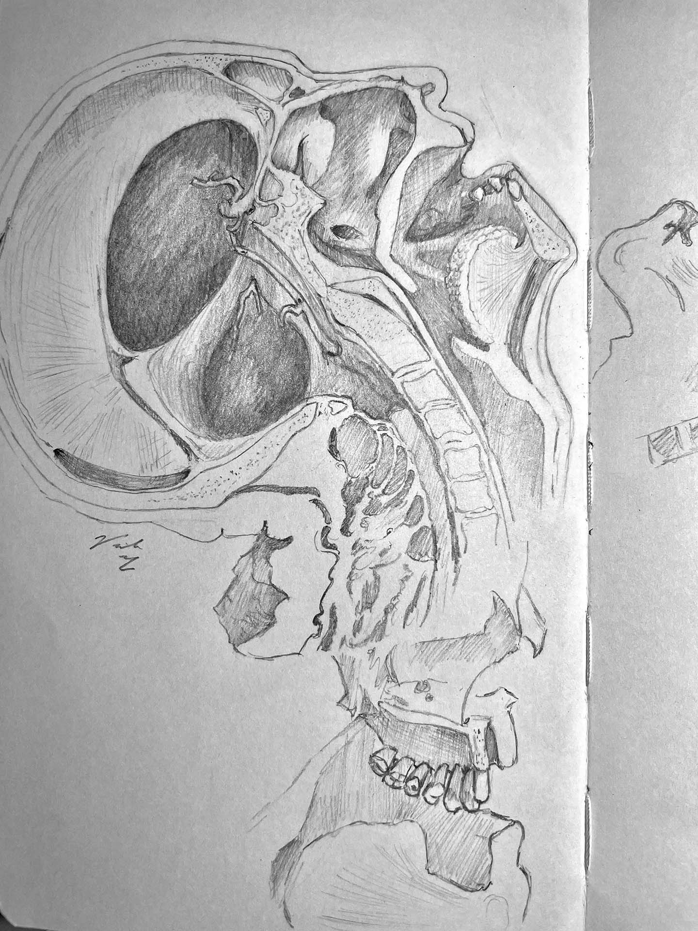
Observational Sketches
At the JCB Grant Anatomy Museum at University of Toronto, there are is a collection
of perserved human specimen. These specimens were used as reference for "Grant's Atlas of Anatomy".
To respect the deceased, no photographs are allowed of the specimens. In fact, the museum is monitored
during all open hours of operations, and all devices in the room must cover their cameras.
For this assignment, we were to use direct observation to illustrate a tonal black and white represenation
of one of specimens and refine it so it looks like living tissue. Therefore, one of the challenges was
reverse engineering the clearly dead and perserved structures to look as it did during life.
For this specimen, it included replacing the missing teeth, repositioning the head upright, and reconstructing
the tissue to look more alive.
